buckle fracture wrist healing time
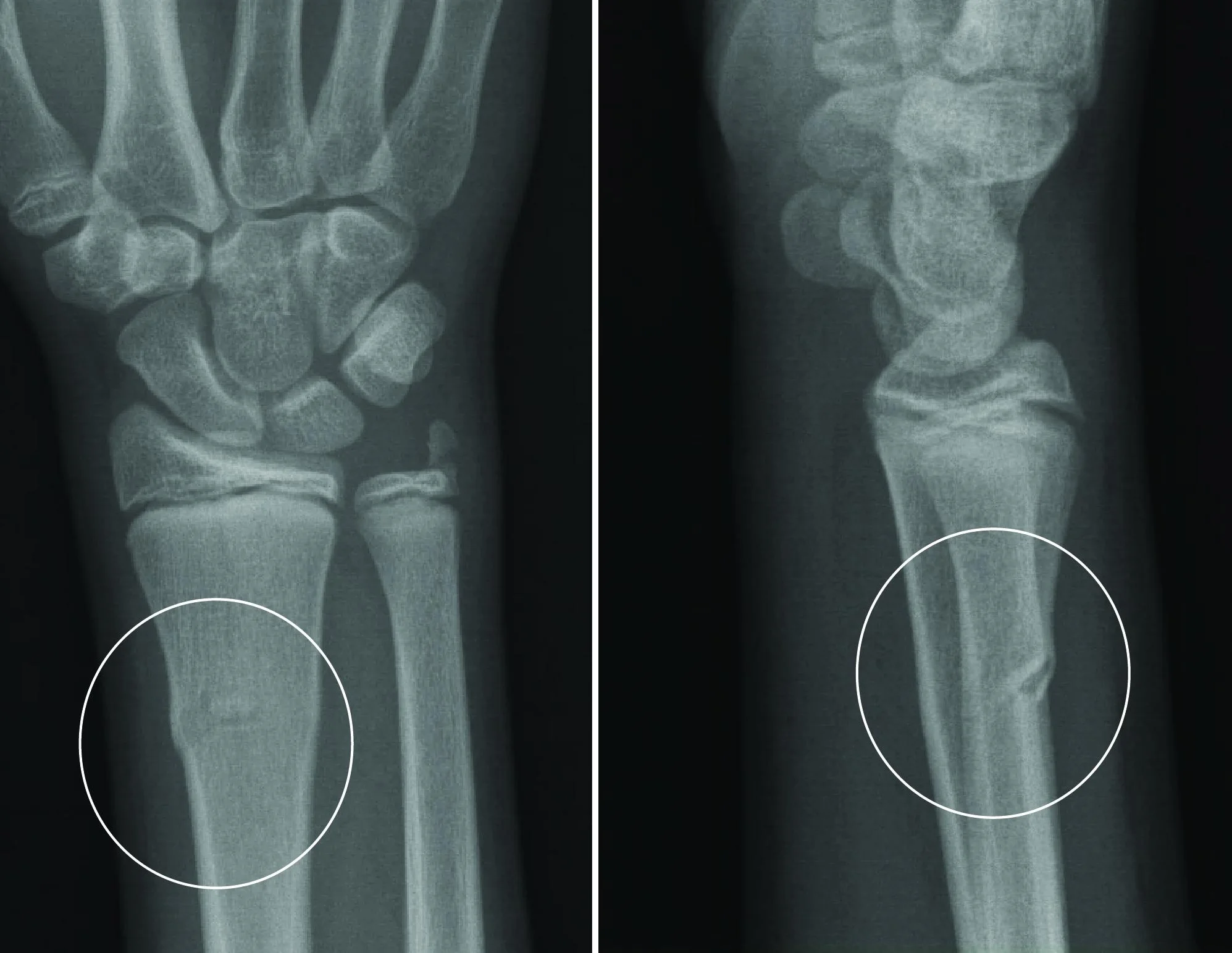 Buckle Fracture - Raleigh Hand Surgery — Joseph J. Schreiber, MD
Singapore Bone Fractures Specialist Centre Get professional treatments and tips on your bone fracture today. Call us at (65) 64712744 or SMS at (65) 92357641 to schedule an appointment with the bonus specialist. is available. A bone fracture is a medical condition in which there is a breakdown in bone continuity. The bones form the skeleton of the body and allow the body to be supported against gravity and move and function in the world. Bones also protect some parts of the body, and the bone marrow is the production center of blood products. Bone FractureBone is not a stagnant organ. It is the body's calcium reservoir and is always experiencing changes under the influence of hormones. Parathyroid hormone increases calcium levels in the blood by healing the calcium of the bone, while calcitonin has the opposite effect, allowing the bone to accept the calcium of the blood. What causes a fracture? When external forces apply to the bone has the potential to fail. Fractures occur when the bone cannot stand those external forces. Fracture, break or crack all means the same. A term is not better or worse than another. The integrity of the bone has been lost and the bone structure fails. Broken bones injured by a variety of reasons, including: Often a fracture is easy to detect because there is obvious deformity. However, it is sometimes not diagnosed easily. It is important that the doctor take a history of the injury to decide which potential problems might exist. In addition, fractures do not always occur in isolation, and there may be associated injuries that need to be addressed. Fractures may occur due to direct strokes, twisted injuries or falls. The type of forces in the bone can determine what type of injury occurs. The descriptions of fractures can be confusing. They are based on:The first step in describing a fracture is whether it is open or closed. If the skin over the fracture is interrupted, then there is an open fracture. The skin can be cut, torn or abracidated (scrated), but if the integrity of the skin is damaged, there is the potential of an infection to enter the bone. Since the bone fracture site communicates with the outside world, these injuries must be aggressively cleaned and often require anesthesia in the operating room to do the job effectively. Then there must be a description of the fracture line. Does the fracture line cross the bone (transverse), at an angle (oblique) or spiral? Is the fracture in two pieces or is it comminus in multiple pieces? Finally, the alignment of the fracture is described as to whether the fracture fragments move or in their normal anatomical position. If bone fragments are not in the right place, they need to be reduced or placed back in their normal alignment. What common types of fractures? Stress fracture A stress fracture is an excessive injury. Due to repeated microtrauma, the bone may not absorb the shock that is being put on it and weakens. Most of the time it is seen in the lower leg, the tibia bone (tibia), or the foot. The athletes are at the highest risk, because they have repeated leaps on hard surfaces. Tennis players, basketball players, jumpers and gymnasts are usually at risk. A March fracture is the name given to a stress fracture of the metatarsal or long bones of the foot. (It is called because it often happens in soldiers who are forced to march long distances.) Diagnosis is performed by history and physical examination, although at times a bone scan can be performed to confirm the diagnosis. Treatment is conservative, rest, ice, and anti-inflammatory drugs such as ibuprofen. These fractures may take six to eight weeks to heal (always the fracture is seen in the X-ray). Trying to get back too fast can cause rejuzation, and can also allow the stress fracture to spread across the bone. Fragments of the tibia may have very similar symptoms such as a stress fracture, but they are due to inflammation of the lining of the bone, called the periosteum. Fragments are caused by overuse, especially in runners, walkers, dancers, including those who make aerobics. The muscles that go through the periosteum and the bone itself can also inflate. Treatment is similar to a stress fracture and physical therapy can be helpful. Compression Fraction As people age, there is a potential for bones to develop osteoporosis, a condition where bones lose their calcium content. This makes the bone more susceptible to breaking. One of these types of lesion is a compression fracture in the spine, most of the time the spinal spine or lumbar spine. Since we are a righteous animal, if the bones of the back are weaker than the force of gravity these bones can be crude. Pain is the main complaint, especially with the movement. The compression lesions of the back may or may not be associated with nerve injury or spinal cord. An X-ray of the back can reveal bone injury, however, sometimes a CT scan or an MRI will be used to ensure that the spinal cord is not damaged. Treatment includes pain medication and often a back brake. Some compression fractures can also be treated with vertebroplasty. Verbroplasty involves inserting a material similar to glue in the center of the collapsed spinal vertebrae to stabilize and strengthen the crushed bone. The glue (methylmethacrylate) is inserted with a needle and syringe through the anesthetized skin in the mediaportion of the vertebra under the guidance of specialized X-ray equipment. Once inserted, the glue soon hardens, forming a structure similar to plaster with the broken bone locally. Fraction of the rib The ribs are especially vulnerable to injury and are prone to breaking due to a direct blow. Rib X-rays are rarely taken as it doesn't matter if the rib is broken or just curled. A chest X-ray is usually taken to ensure that there is no collapse or hematoma of the lung. When we breathe, it's like a serene. We inhaled the air in our lungs and the ribs move out and the diaphragm moves down. When a person has a rib injury, the pain associated with it makes breathing difficult, and the person has a tendency not to breathe deeply. If the underlying lung of the lesion does not expand, it is at risk of infection. The person is then susceptible to pneumonia (pulmonary infection), which is characterized by fever, cough and shortness of breath. Unlike other parts of the body that can rest when injured, it is very important to take deep breaths to prevent pneumonia when rib fractures are present. The treatment for the hermetic and broken ribs is the same: ice on the chest wall, ibuprofen as anti-inflammatory, deep breathing and analgesic. With lower rib fractures, there may be concern about the organs of the abdomen that protect the ribs. The liver is below the ribs on the right side of the chest, and the spleen under the ribs on the left side of the chest. Many times your doctor may be more concerned about the abdominal injury than the broken rib itself. Ultrasound or CT scan can help diagnose intraabdominal lesions. Skull fracture With the wide availability of CT scans, X-rays of the skull are rarely taken to diagnose head injuries. If there is a head injury, the doctor will feel or palp the scalp and skull to determine whether there can be a fracture in the skull. You will also look in the ears to see if there is blood behind the ear drum and also complete a neurological examination. The skull is a flat, compact bone and requires significant force to break it. If there is a cranial fracture, there is a greater probability of bleeding in the brain, especially in children. There are guidelines available to decide whether to indicate a CT scan (needed). The minor head injury is defined as loss of consciousness, defined amnesia or presence in patients with a GCS score (Glasgow Coma Score) of 13-15. With minor injuries to the head, the following risk groups are considered when assessing the need for a CT scan: High risk for neurosurgical potential Media risk (for brain injury in the CT)DiagnosisThe diagnosis of bone fracture is performed by the doctor when asking the patient initially the cause of the injury, when the injury occurred and what other part of the body is suffering. X-rays will be taken if the doctor sees the symptoms of bone fracture through the physical exam. If there is a complete fracture with total bone dislocation and fragmented bones, this injury will be considered as an emergency and will require surgery and internal fixation. Some doctors directly recommend the use of MR or TC to see clearer images of the bones and thus even fractures in the hair can be detected. If the patient suffers from osteoporosis, scans or X-rays are also recommended to know the total coverage of the fracture. Rehabilitation or rehabilitation is necessary. Treatment process: There are also different ways to treat bone lesions, but the most popular type is the use of plaster especially if the injured part is anywhere in the limb. The bones that move will be put back in place with the use of bone traction. No surgery is needed if there are no bone fragments that will be revealed by the X-rays. The plaster will immobilize the extremity until the bones heal. The person will have to undergo physical therapy when the bone has finally joined again but the person should avoid heavy stress to prevent the recurrence of the fracture. The second option is surgery and the use of internal fixing. This is done for complete fractures with multiple fragments. The use of wires, pins and plates is inevitable because there is a need to readjust the bones completely. For long bones, metal bars are inserted through the bones to unite the bones. External fixing can also be used to correctly align the long bones. In this type of treatment, long bones are connected with metal pins and these pins are attached to the external fixer that is placed outside the skin on the injured area. The external fixer is also made up of metal rods with adjusters and is there to prevent the bones from coming out of position until it heals. How the treatment works: In the internal fixing, when the bone is fixed through wires, pins or metal rods, the bone will produce calluses inside the broken part once the bones are joined. These calluses harden in calcium deposits and close the cracks completely. The same is true of the bones that were subjected to reduction or bone melting. Bone healing time or recovery time can be from weeks to months depending on the severity of the fracture and if there are no complications that followed the treatment. Physical therapy continues after the bone has been removed from its plaster. This is to strengthen muscles and condition the bone so that bone remodeling can take place. Prevention There are many ways to prevent fractured bones from happening but the best way is to avoid doing anything that can cause damage to your body. In sports, there are protective gears that you could use to protect the bony parts of your body, such as knee pads, wrist bands, elbow pads, helmets, and enamel pads and so on. Although these things can't stop the injury completely, at least the risk of having a severe fracture can be minimized. about the stress fracture on the internal fixing of open reduction (ORIF) Surgery on Body Fracture in Children on Surgery for RotoMake Bonus Fracture Appointment or Question: Call: (65) 64712744 / Whatsapp or SMS: (65) 92357641 / Email: info@boneclinic.com.sg – 24 hours HotlineMake Appointment or Question:Call: (65) 64712744 / Whatsapp or SMS: (65) 92357641 / Email: info@boneclinic.com.sg – 24 hours HotlineContact Details

OrthoKids - Forearm Fractures

Fracture: How to treat a buckle fracture of the distal radius
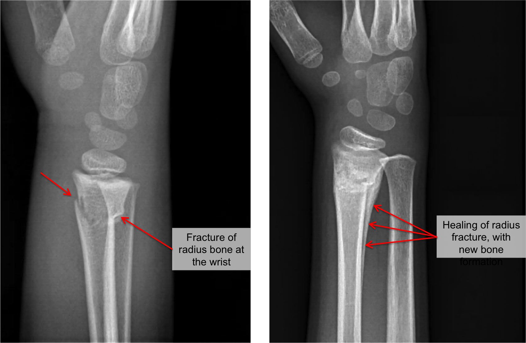
Buckle Fracture - Raleigh Hand Surgery — Joseph J. Schreiber, MD
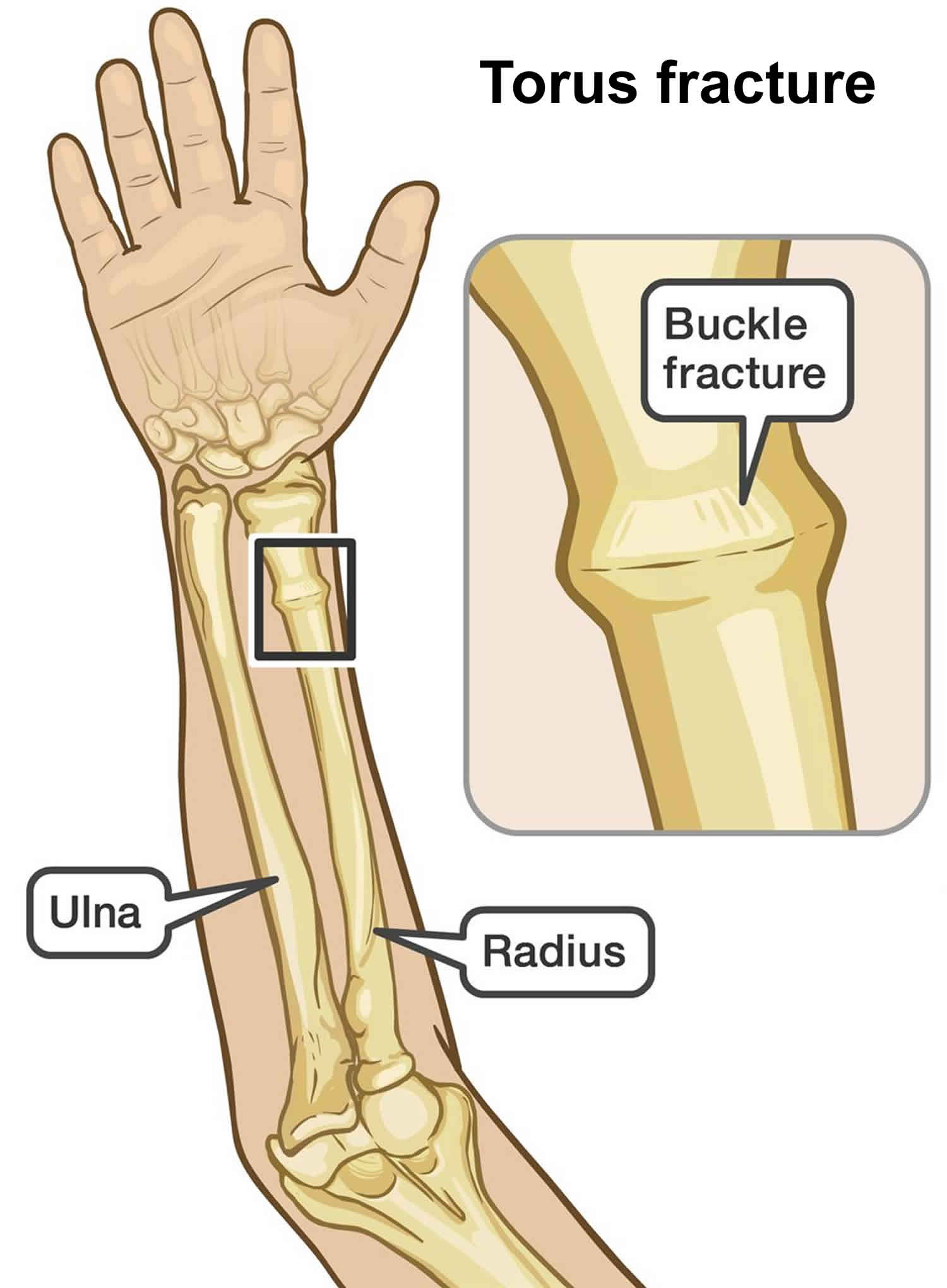
Buckle fracture causes, symptoms, diagnosis & treatment
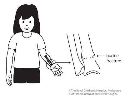
Kids Health Information : Fracture care: buckle injury

OrthoKids - Forearm Fractures

Buckle Fracture - Raleigh Hand Surgery — Joseph J. Schreiber, MD

Buckle (Torus) Fracture of an Arm

OrthoKids - Forearm Fractures
Choosing Wisely: Distal Radius Buckle Fractures - CanadiEM
Buckle Fracture by Dr. David Nelson, MD
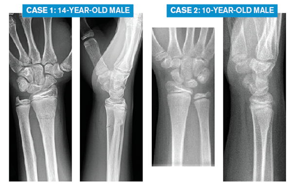
Emergency Medicine Pearls, Pitfalls for Treatment of Pediatric Distal Radius Fractures - ACEP Now

What are Buckle Fractures? - WristSupports.co.uk
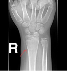
The Pediatric Wrist Buckle Fracture is Common - Louisville Bones
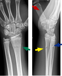
Emergency Medicine Pearls, Pitfalls for Treatment of Pediatric Distal Radius Fractures - ACEP Now

Fractures - Distal forearm or wrist
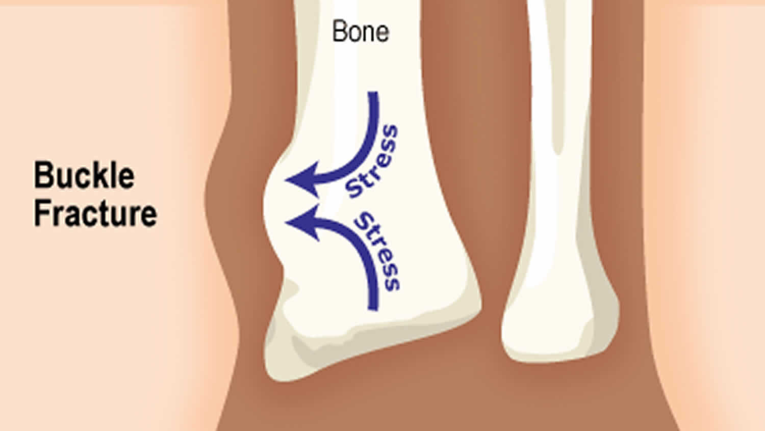
Buckle fracture causes, symptoms, diagnosis & treatment

What level of immobilisation is necessary for treatment of torus (buckle) fractures of the distal radius in children? | The BMJ
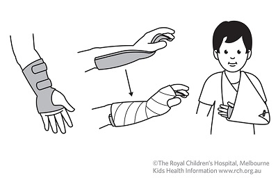
Kids Health Information : Fracture care: buckle injury

Distal radius fracture - Wikipedia
OrthoKids - Forearm Fractures
Forearm Fractures in Children - Types and Treatments - OrthoInfo - AAOS
A New "Rule" for Differentiating Stable Buckle and Other Distal Radius Fractures

MedPix Case - Distal Radius Torus Fracture/Buckle Fracture
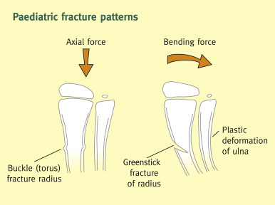
Greenstick or Buckle Fracture - What Are They & How to Get Treatment

Wrist buckle fracture | Sydney Children's Hospitals Network
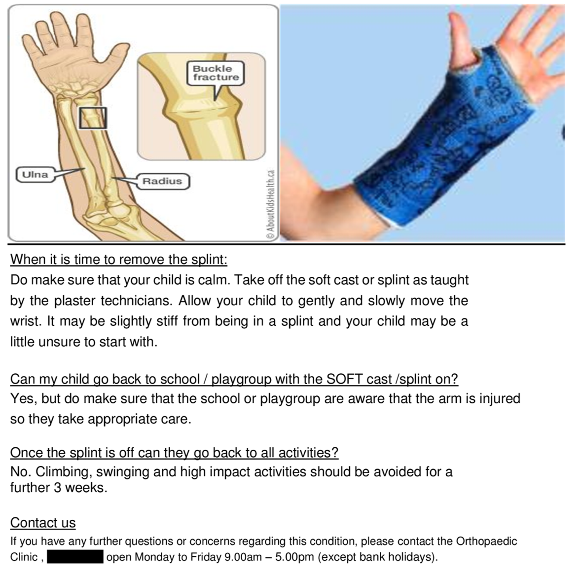
Cureus | Improving Management of Paediatric Buckle Fracture in Orthopaedic Outpatients: A Completed Audit Loop

Fracture: How to treat a buckle fracture of the distal radius
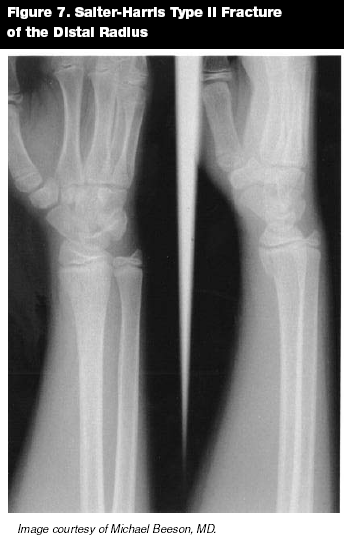
Scooter Fractures, Buckle Fractures, and Beyond: Pediatric Hand and Wrist Injuries in the ED | 2005-08-01 | AHC Media: Continuing Medical Education Publishing
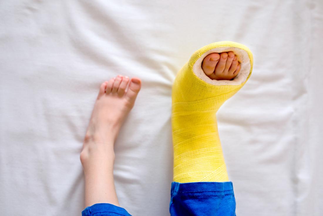
Buckle fractures: Risk factors and recovery

OrthoKids - Forearm Fractures
Buckle fracture of wrist

Pediatric Distal Forearm and Wrist Injury: An Imaging Review | RadioGraphics

Fractures | Boston Children's Hospital

Buckle Fracture: Causes, Recovery, and More
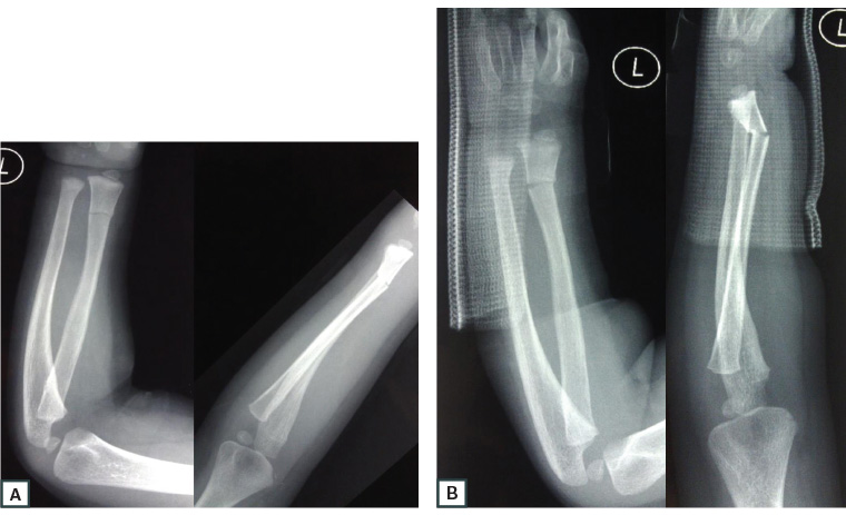
RACGP - Guide to paediatric forearm fractures

Wrist Fracture - Open Reduction and Internal or External Fixation - Patient Education | Colonial Healthcare
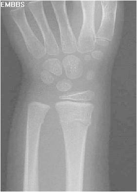
Distal Radius Fractures - Pediatric - Pediatrics - Orthobullets

Buckle fractures: Risk factors and recovery
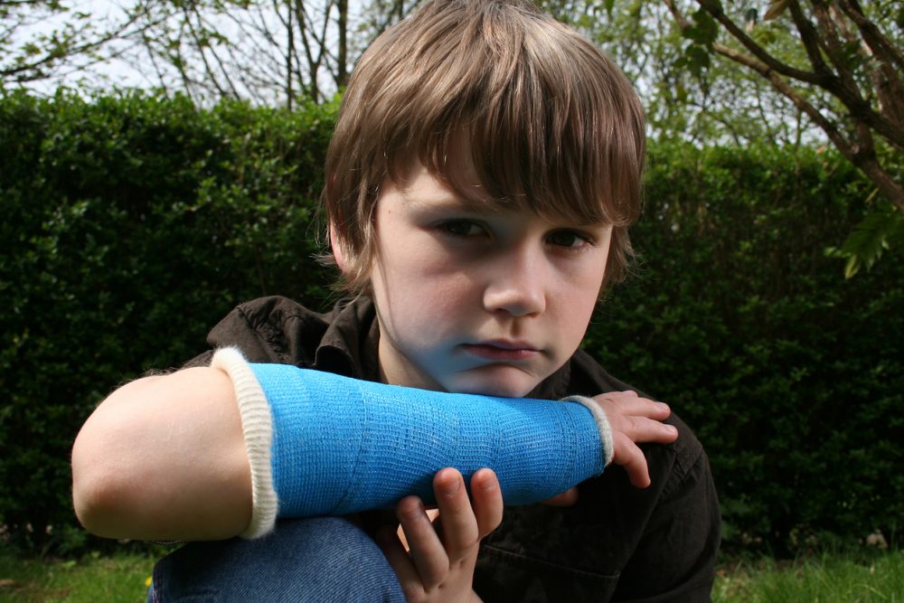
Wrist fractures in children: how are they best treated? - Evidently Cochrane
Posting Komentar untuk "buckle fracture wrist healing time"